Cryo-electron microscopy (cryo-EM) has become a cornerstone technique in structural biology, offering powerful insights into macromolecular complexes and cellular structures in their native environments. With the advent of highly stable high-resolution microscopes, direct electron detectors, and automated data collection systems, researchers can now routinely resolve biomolecular structures at near-atomic resolution using both single-particle and tomography methods. Atomic models can be built directly into cryo-EM maps, revealing precise molecular interactions and functional conformations. Additionally, hybrid approaches that combine cryo-EM with optical and scanning electron microscopy enable detailed visualization of targeted cellular structures. This versatility—spanning molecular to cellular scales—makes cryo-EM a vital tool in both structural and cell biology. It is especially useful for dissecting dynamic macromolecular machines by capturing their various conformational states. This EMBO Practical Course is designed to provide participants with a solid foundation in image processing for cryo-EM, focusing on high-resolution data analysis for both single-particle and tomographic datasets. Aimed at advanced PhD students and postdocs, the course offers practical training for those using cryo-EM to pursue structural investigations.

This event has passed.
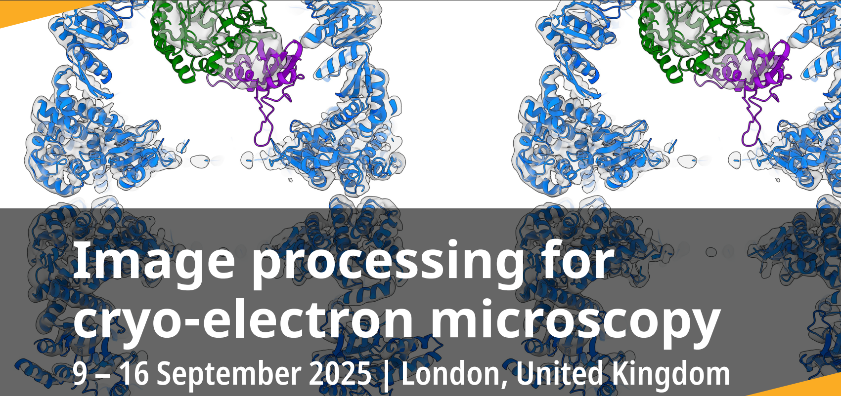
Image processing for cryo-electron microscopy
September 9 @ 2:00 pm - September 16 @ 3:00 pm